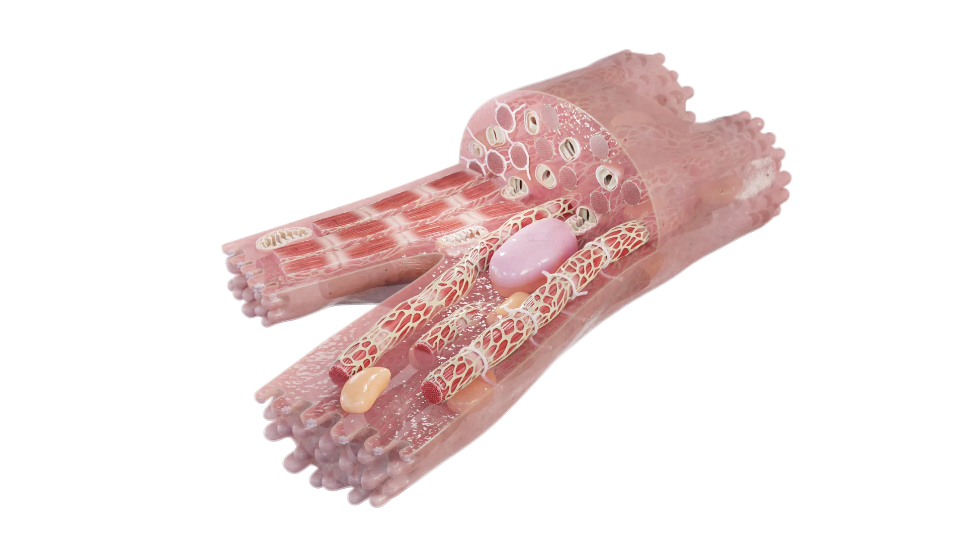
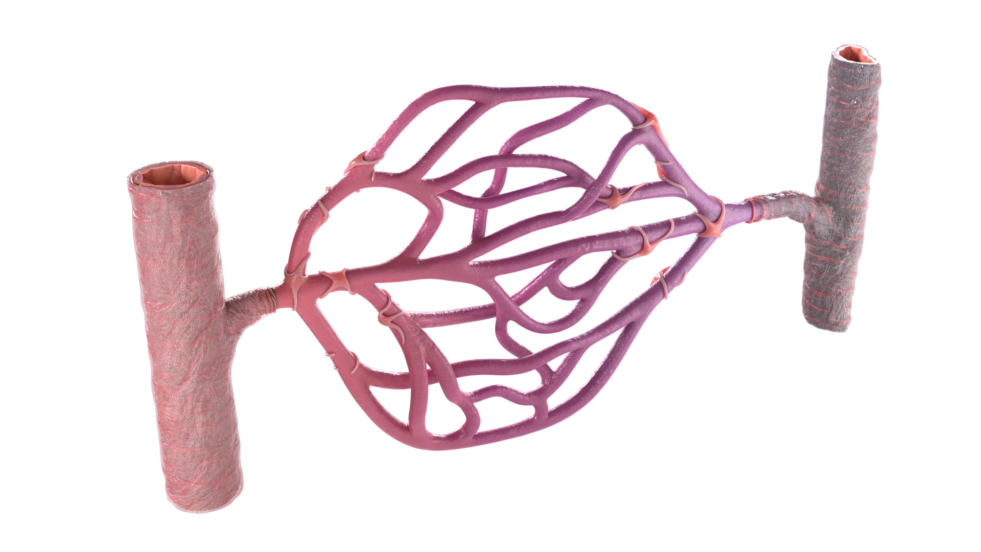
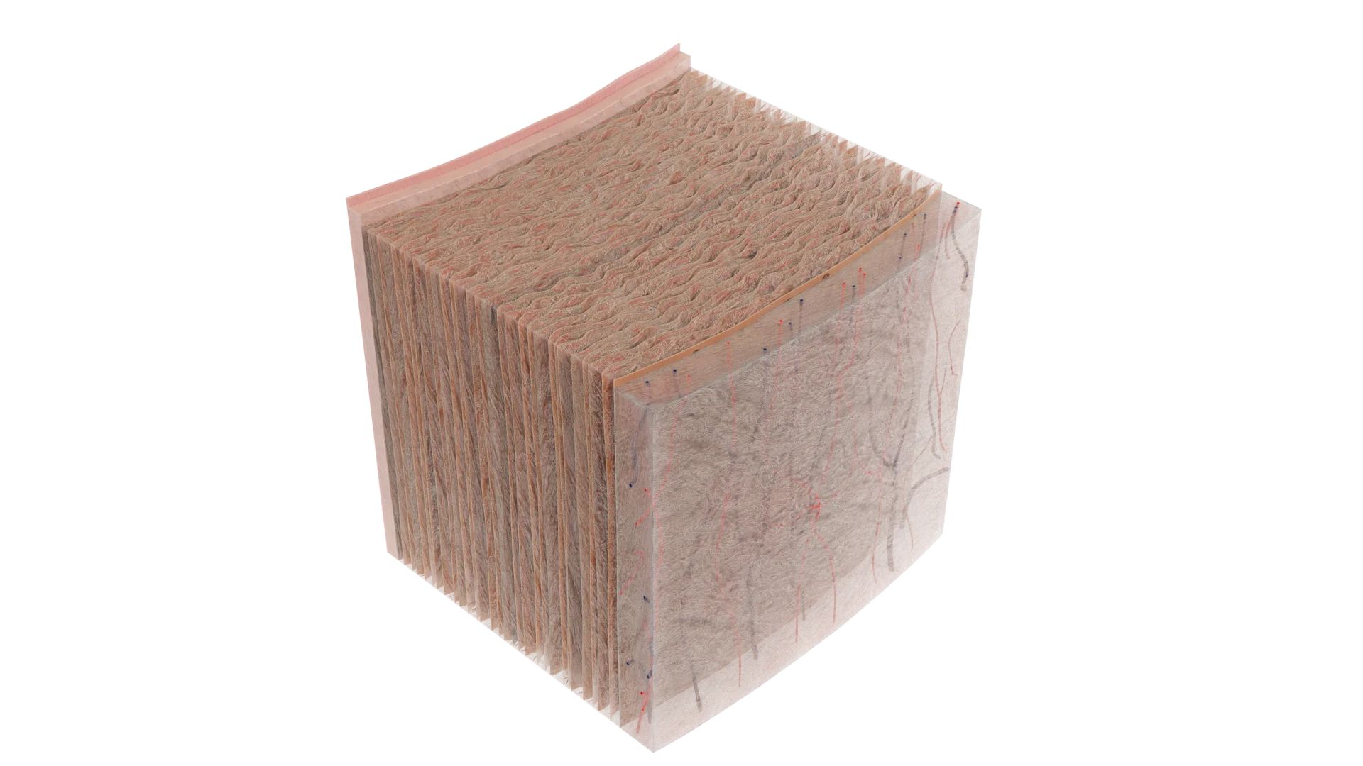
Explore a 3D microanatomy model of the human circulatory system at the microscopic level, revealing the intricate pathways of blood flow through tissues and cells. Rotate, zoom, and dissect layers to examine cardiac myocytes, endothelial cells, valve structures, and the microarchitecture of arterioles, venules, and capillaries. Every component—from vessel walls to cellular junctions—is meticulously labeled, transforming the model into a dynamic learning tool for students, educators, and medical professionals studying histology and vascular physiology.
Show more Show less

Fill out the form and we’ll get back to you once we’ve processed your request.
You can contact us using: