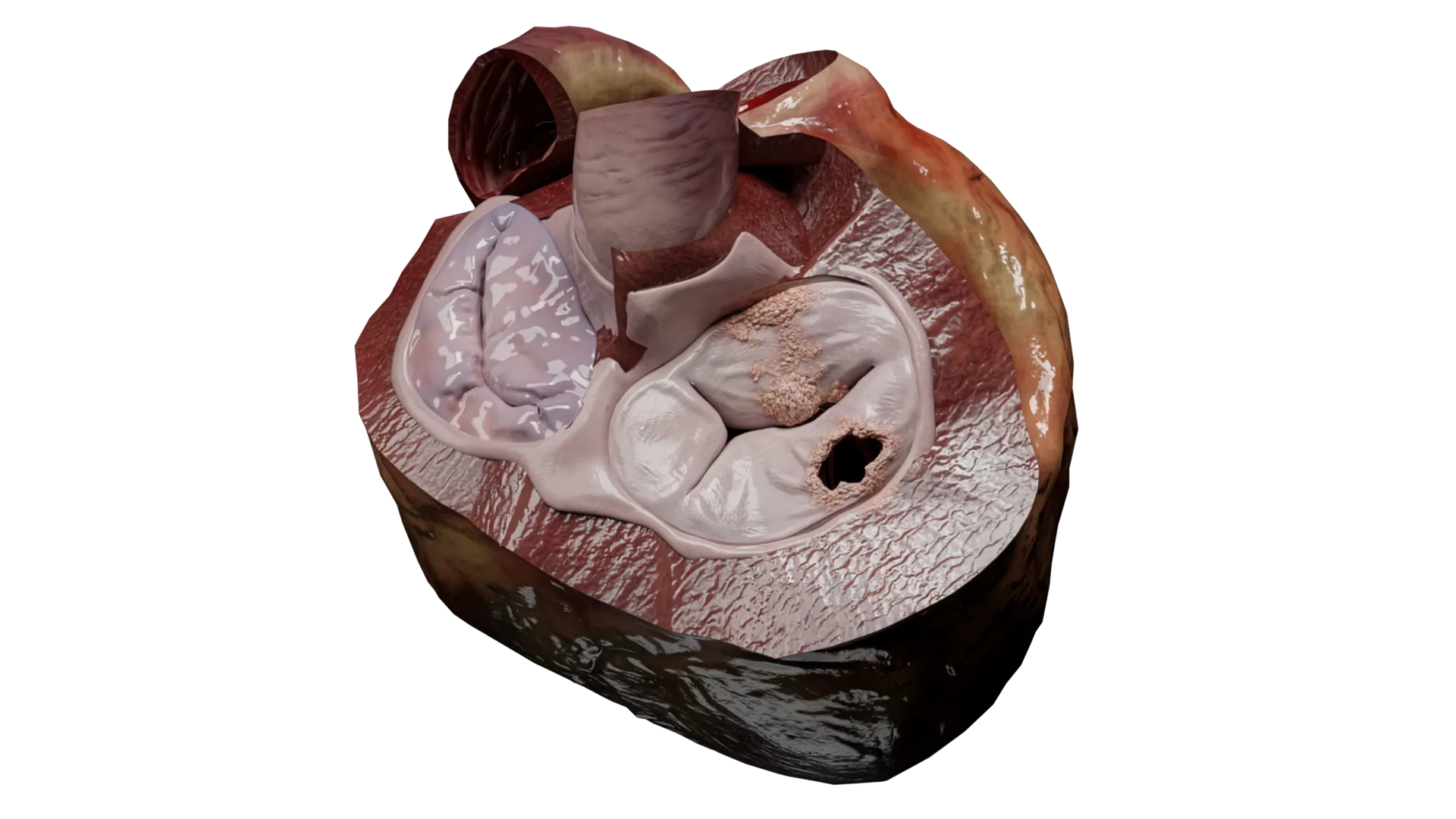
Visualize the features of a left ventricular pseudoaneurysm after myocardial infarction with this 3D model. It highlights a contained rupture of the ventricular wall, bounded by pericardium or scar tissue, distinguishing it from a true aneurysm. The model helps learners and clinicians understand the high risk of rupture, altered hemodynamics, and the urgent need for surgical repair. Its interactive representation supports clinical education, imaging interpretation, and preoperative planning.
Show more Show less