Browse a collection of multi-purpose 3D medical short videos created for marketing teams, educators, clinicians, and patients or explore our 3D medical animation services
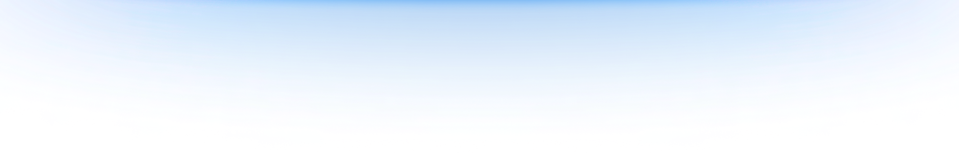
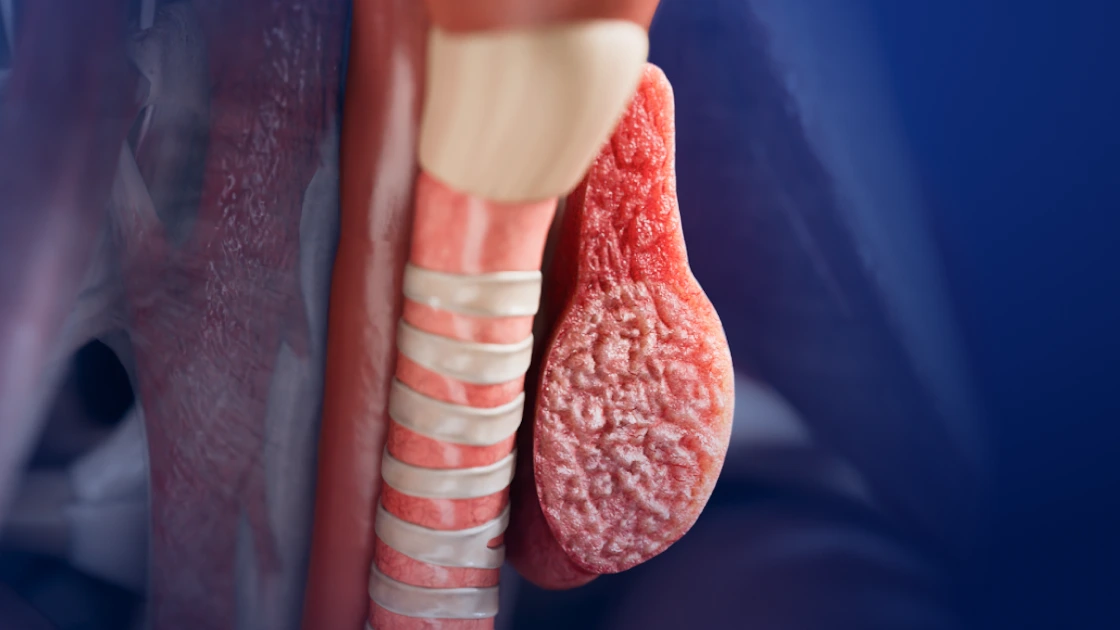
Endocrinology
Shorts
Multinodular goiter

Shorts
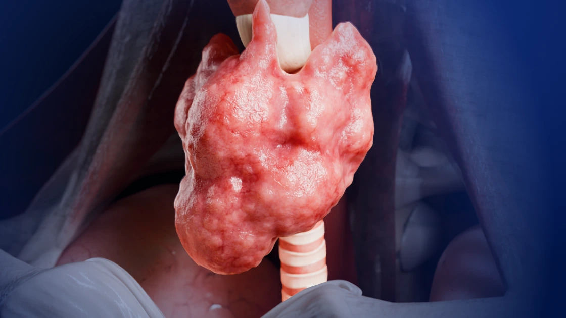
Traumatology
U-shaped sacral fracture
Shorts
Traumatology
Zona 1 sacral fracture
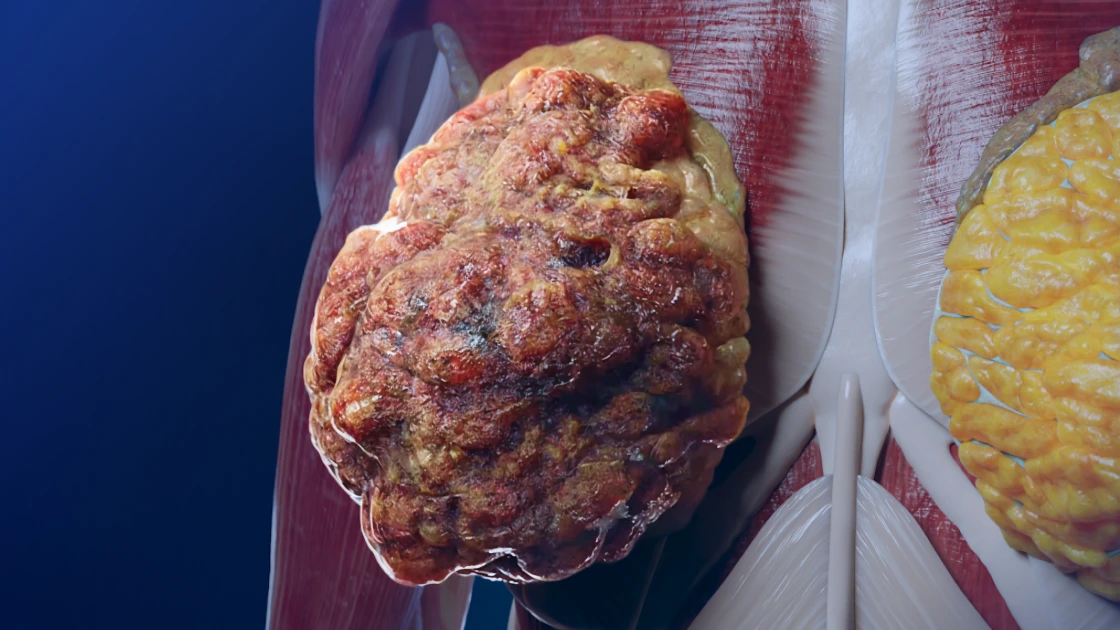
Oncology
Shorts
Invasive lobular carcinoma
Endocrinology
Shorts
Nodular goiter

Endocrinology
Shorts
Diffuse goiter
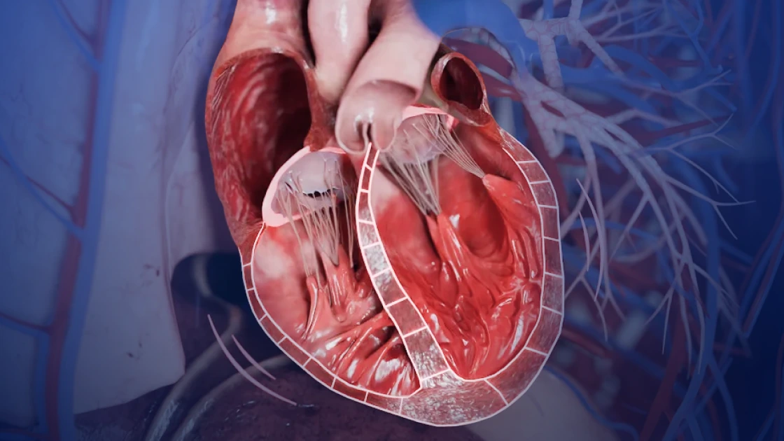
Cardiology
Shorts
Dilated cardiomyopathy
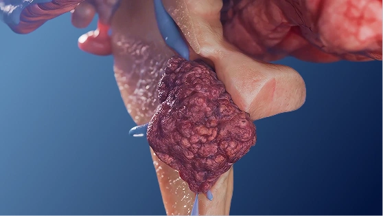
Oncology
Shorts
Ependymoma
Oncology
Shorts
High-grade Fallot’s tumor
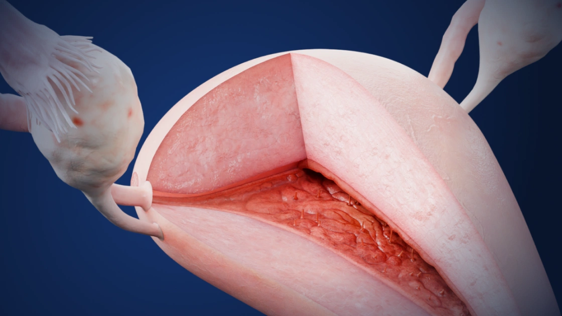
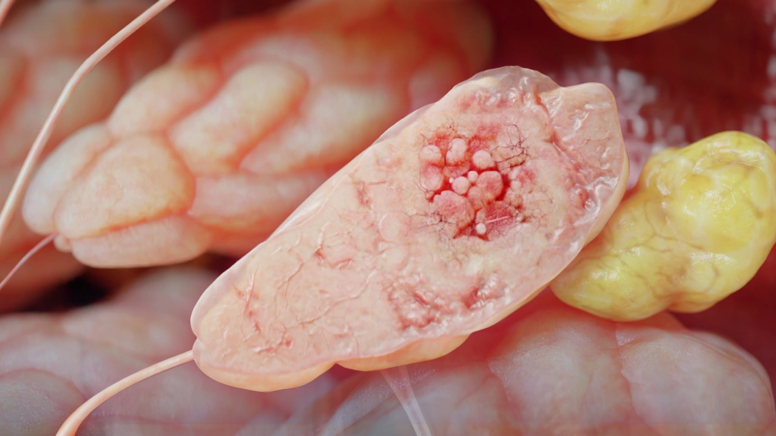
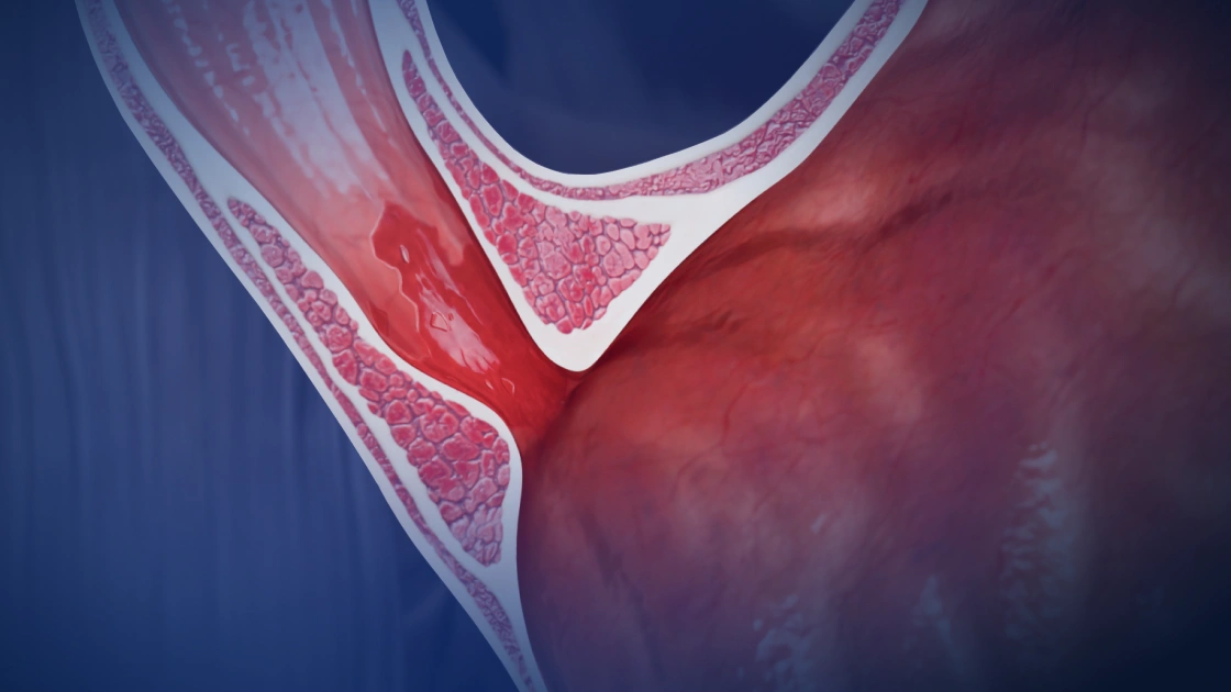
Gynecology
Shorts
Tuberculous endometritis
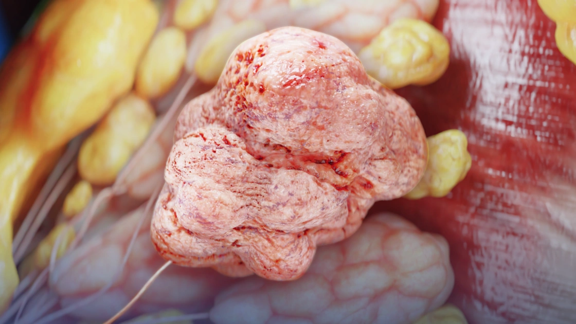
Oncology
Shorts
Lobular carcinoma in situ
Gastroenterology
Shorts
Barrett’s esophagus